Figure 4.
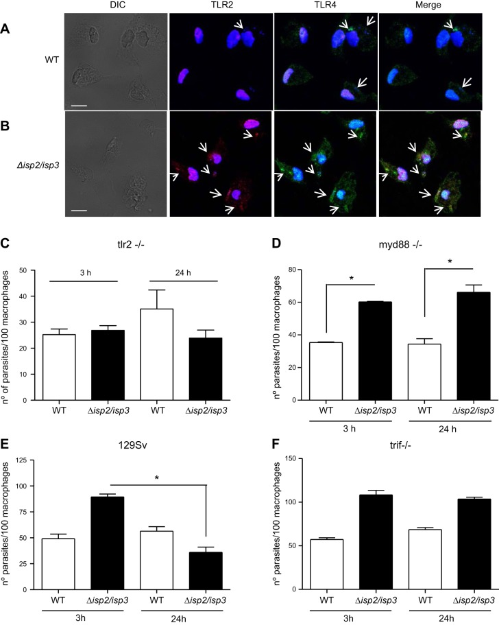
TLR2 and TLR4 are recruited to the site of entry of Δisp2/isp3 L. major in macrophages, and parasite killing requires MyD88 and TRIF. A, B) Macrophages of C57B6 mice were infected with WT (A) or Δisp2/isp3 L. major (B) for 1 h and processed for immunofluorescence, with anti-TLR2 (red) or anti-TLR4 (green). Samples were analyzed by confocal microscopy, sectioned from bottom to top at 0.9 μm, 8 times; images show section 3. Scale bars = 5 μm. Arrows indicate the parasite. C–F) Macrophages from TLR2-knockout mice (C), MyD88-knockout mice (D), 129Sv mice (E), or TRIF-knockout mice (F) were infected for 3 h, washed, and fixed or cultivated for a further 24 h. Experiments were performed 2 separate times. *P < 0.001.

