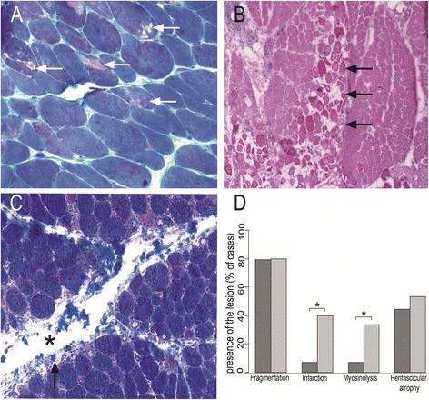
Figure 2.

Comparison of myopathological lesions in DM (A, B) and ASM(C). A: Myosinolysis defined by the presence of punched-out vacuoles (arrows) or myofibrillar rarefaction areas, corresponding to foci of myosin filament proteolysis. B: Microinfarcts defined by numerous contiguous fibres within a fascicle, at the same stage of necrosis or regeneration. C: Perimysial fragmentation defined by focal clear areas (asterisk) and spumous appearance of adjacent areas (arrowhead). D: Percentages of cases with presence of the lesion (perimysial fragmentation, microinfarcts, vacuoles, and perifascicular atrophy) in ASM and DM. (*) indicates p < 0.05.Frozen sections (A-C); hematoxylin-eosin (A, B) and Masson trichrome (C).
