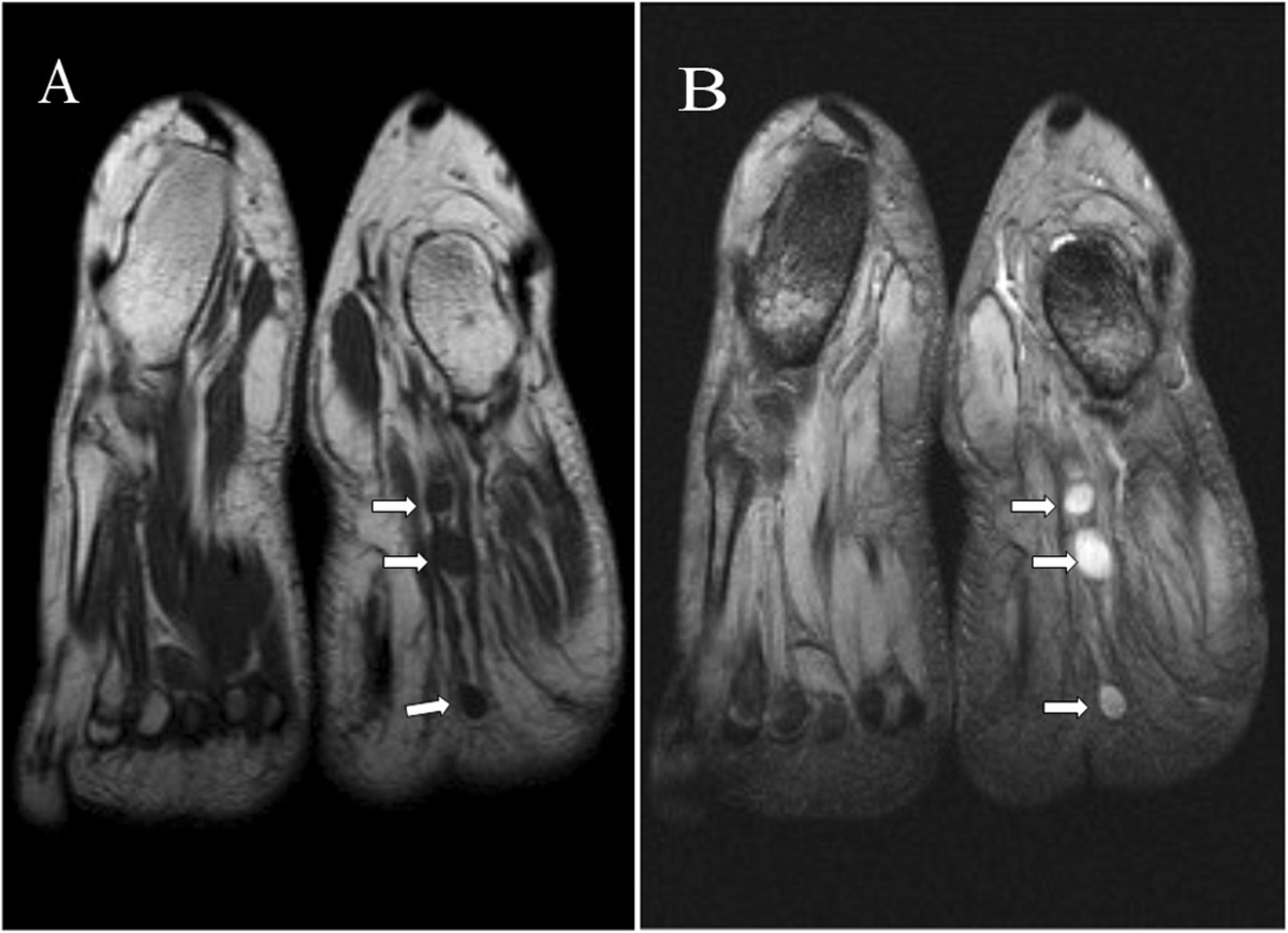
Figure 1.

Coronal MR images showing linear round lesions. (A): T1-weighted fast-spin echo image (TR/TE: 512/11) shows the lesions with homogeneous isointensity to skeletal muscle (arrows). (B): T2-weighted multi echo data image combination sequence (TR/TE: 865/23) demonstrates three prominent nodules of high intensity signal (arrows).
