
Figure 2.
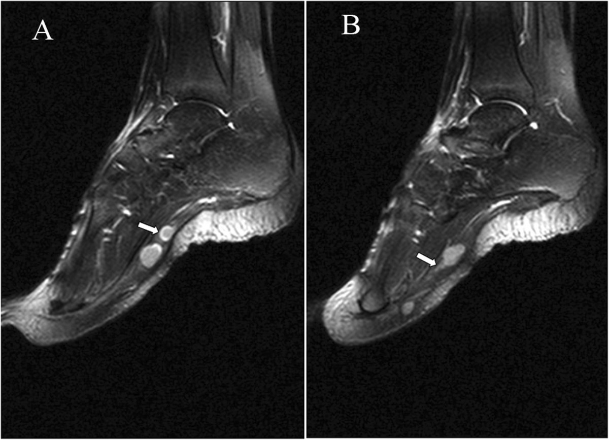
Sagittal T2 fast spin-echo-weighted fat-suppressed image (TR/TE: 3000/40, TI: 150) shows nodules localized in the plantar aspect of the foot. (A): The rim of the tumor shows hyperintense signal in comparison with inside of the tumor (arrow). (B): The surrounding nerve seems like a mouse-tail (arrow).