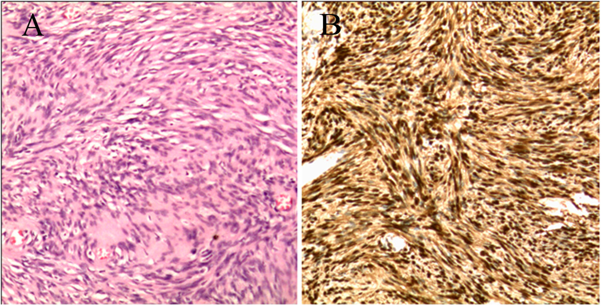
Figure 4.

Photomicrograph of the tumor. (A): Microscopic specimen of the lesion shows long spindle-shaped tumor cells and nuclei palisading with multiple fascicles (Hematoxylin-eosin, H & E, original magnification × 200). (B): The tumor cells stain positive for S-100 protein and exhibit strong immunoreactivity for S-100 protein (Original magnification × 200).
