
Figure 4.
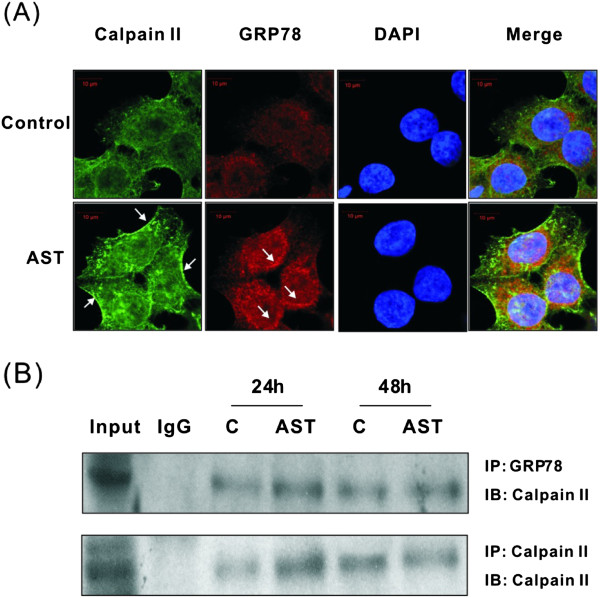
Effects of AST on the localization of (A) and interaction between calpain II and GRP78 (B) were determined. HCT116 cells were treated with AST (80 μg/ml) for 24 h. Co-localization of calpain II and GRP78 were detected in control [C] and drug-treated cells. Specific immunofluorescence antibodies against calpain II and GRP78 were used, while DAPI acted as nuclear stain. Green and red fluorescence indicates the presence of calpain and GRP78, respectively in the cells and blue color represents the DAPI-stained nucleus. Interaction between calpain II and GRP78 was further detected by immunoprecipitation [IP]. HCT116 cells were treated with AST (80 μg/ml) for 24 or 48 h. Immunoprecipitated proteins were collected and subject to Western immunoblotting [IB]. Data shown are representative immunoblots with similar findings. IgG was used as precipitating antibody in the negative control.