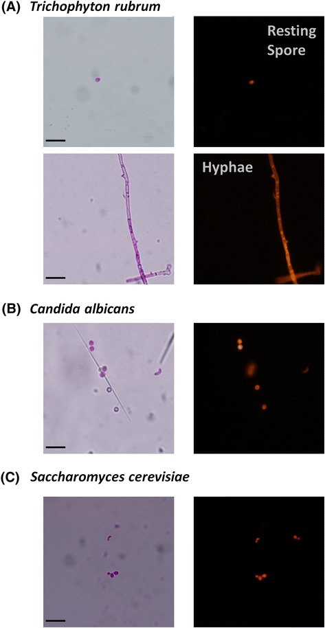
Figure 3.

Micrographs showing the uptake of Rose Bengal by fungi (A) resting spores and hyphae of T. rubrum , (B) C. albicans and (C) S. cerevisiae . The images were captured under brightfield and fluorescence microscopy at 400 X magnification; the scale bar (Bottom right) is 10 μm.
