
Figure 5.
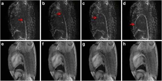
In vivo left heart catheterization positive contrast and anatomical images from dual echo pulse sequence. Positive contrast (a-d) and anatomical images (e-h) generated by the dual echo sequence when the tip is descending aorta (a, e), at the aortic arch (b, f), at the aortic valve (c, g) and in the left ventricle (d, h). Red arrows are used to indicate the wire tip.