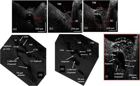
Representative two-dimensional (2-D) structural OCT and scanning electron microscopy (SEM) images from the limbal region of an eye. (a) shows the OCT image captured with the cannula inside SC (arrow). At the location
away from the cannula tip, (b) shows the tissue before infusion of perfusate where arrows identify the TM and SC, while (c) shows the maximally dilated appearance of the same segment. Please also see Video
1 (MOV, 3.63 MB) [URL:
http://dx.doi.org/10.1117/1.JBO.19.10.106013.1]. (d) and (e) are representative SEM images from the limbal region, illustrating structural features of the outflow system that are mirrored in both the SEM and OCT images. Original SEM images:
magnification. (d) shows a collector channel entrance or ostia (CCO). A septum is present at the CCO that is attached to the TM by cylindrical attachment structures (CAS). (e) is the adjacent section from the same segment showing the transition from the region of a CCO in (d) to a circumferentially oriented collector channel. (f) The
enlargement OCT image that is cropped from (c) permits a more detailed comparison of relationships. CM, ciliary muscle.

