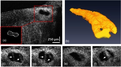
Fig. 4.

(a) A representative B-scan with the region of interest, containing SC (red box), and the segmented SC from the same B-scan is shown in the inset. (b) Side view of the reconstructed 3-D SC. (c) to (f) Selected cross-sections from the SC showing its various shapes along a 2 mm length of a limbal segment. White arrows indicate locations of cylindrical attachment structures spanning between the TM and SC external wall.
