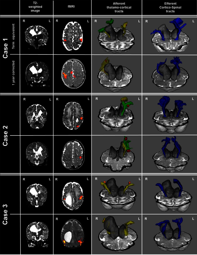
Fig. 2.
Functional activation and probabilistic tractography in three cases with focal periventricular brain injury, studied at term equivalent and 1-year corrected age. A unilateral periventricular white matter lesion can be seen arising from the lateral ventricle at the site of the previous haemorrhagic infarction on the right (cases 1 and 3) and left (case 2) sides. Following passive sensori-motor stimulation of the contralesional hand, clusters of functional activity were identified in all infants at both time-points in the ipsilesional perirolandic region (z-score threshold 2.3). The afferent thalamo-cortical tracts (yellow and green) developed altered trajectories which circumvented the periventricular white matter lesion to meet the identified fMRI clusters (orange/red). Efferent corticospinal tracts (blue) showed marked asymmetry, with a decreased volume in the lesional hemisphere evident at both time-points

