Fig. 5.
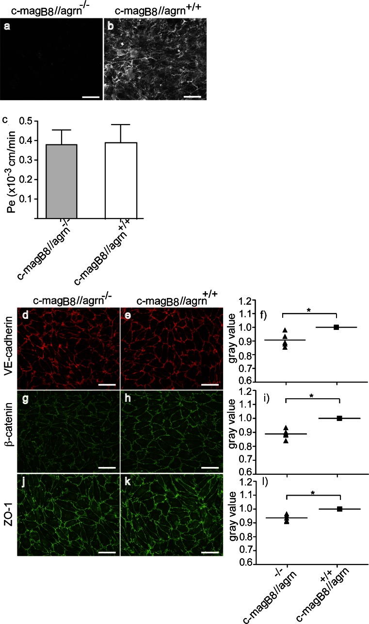
Agrin−/− pMBMECs show reduced junctional localization of VE-cadherin, β-catenin, and ZO-1. a, b Primary mouse brain microvascular endothelial cells (pMBMECs) were isolated from rescued agrin knock-out (c-magB8//agrn−/−) or control littermates (c-magB8//agrn+/+), and after 7 days in culture, immunofluorescence staining for agrin was performed. The presence of the extracellular matrix protein agrin is visible by specific binding of the fluorochrome-labeled anti-agrin antibody in control cells (white signal in b), and the absence of this binding on agrin-knock-out endothelial cells (no fluorescence signal in a). Micrographs from one representative experiment out of five are shown. Bar 50 μm. c Permeability of agrin-deficient (c-magB8//agrn−/−) and control pMBMECs (c-magB8//agrn+/+) was measured after 7 days in culture for 3-kDa Dextran. Bars represent the mean of seven independent experiments (± SEM). d–i After 7 days, pMBMECs isolated from rescued agrin knock-out (c-magB8//agrn−/−) or control littermates (c-magB8//agrn+/+) were stained for VE-cadherin (d, e), β-catenin (g, h), and ZO-1 (j, k), and the junctional IF signal quantified by measuring the gray values at the junctions (f, i, l). Micrographs of one representative experiment out of five are shown. Bars 50 μm. The gray values of the knock-out cells were normalized to value of the control cells, which was set to 1. Each symbol represents the mean gray value of all five independent experiments, and the horizontal lines indicate the means of all experiments performed. *P < 0.05

