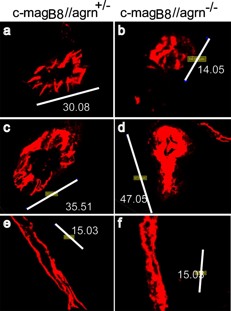
Fig. 6.
Agrin stabilizes junctional localization of VE-cadherin in brain microvascular endothelial cells in vivo. Three-dimensional reconstruction of VE-cadherin (red)-immunostained brain vessels. In each column, vessels from three different mice are shown. In c-magB8//agrn−/− mice (b, d, f), VE-cadherin immunostaining is more diffuse and does not display a clear junctional staining pattern compared with the staining observed in vessels of heterozygous control animals (c-magB8//agrn+/−; a, c, e). Indicated numbers represent the length of the white bar in micrometers. Representative micrographs of three animals out of four agrin knockout and four control animals are shown

