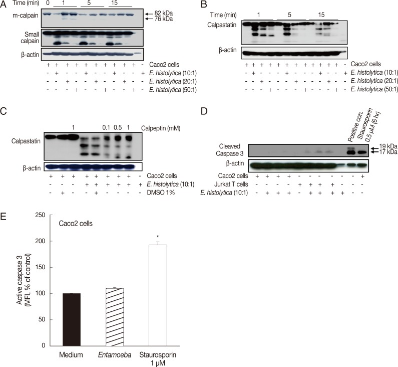
Fig. 1.
Incubation with E. histolytica induces cleavage of calpain and calpastatin, but not caspase-3, in Caco-2 cells. (A, B) Caco-2 cells (1×10⁶/sample) were incubated for 1-15 min at 37℃, either with or without E. histolytica at ratios of 10:1, 20:1, or 50:1 (Caco-2 cells to E. histolytica). After incubation, proteins in whole cell lysates were subjected to SDS-PAGE and then blotted with anti-m-calpain, anti-calpain small subunit, and anti-calpastatin antibodies (Abs). β-actin was used as a loading control. (C) Caco-2 cells (1×10⁶/sample), pretreated either with or without calpeptin, were incubated for 15 min at 37℃ in either the absence or presence of E. histolytica (1×10⁵/sample) in a CO2 incubator. After incubation, proteins in whole cell lysates were subjected to SDS-PAGE and blotted with anti-calpastatin Abs. β-actin was used as a loading control. (D) Caco-2 cells were incubated with E. histolytica (E. histolytica to Caco-2 ratio, 1:10) at 37℃ for 15 min. Proteins in whole cell lysates were analysed for the presence of activated caspase-3 by immunoblotting with anti-cleaved caspase-3 Abs that recognize the p19 and 17 fragments of caspase-3. Medium alone served as a negative control. Caco-2 cells incubated with staurosporin (1 mM for 6 hr) and Jurkat T cells incubated with amoebae served as positive controls. (E) Caco-2 cells were incubated with E. histolytica (E. histolytica to Caco-2 ratio, 1:10) at 37℃ for 20 min and subsequently stained for activated caspase-3 with FITC-conjugated rabbit anti-active caspase-3 monoclonal antibodies. Activation of caspase-3 was analysed by flow cytometry. Caco-2 cells were incubated in medium alone as a negative control. Staurosporin (1 mM for 6 hr), a known inducer of apoptosis, served as a positive control. Significant differences between groups are indicated as follows: *P<0.005. Figures are representative of three independent experiments, each showing similar results.

