Fig. 3.
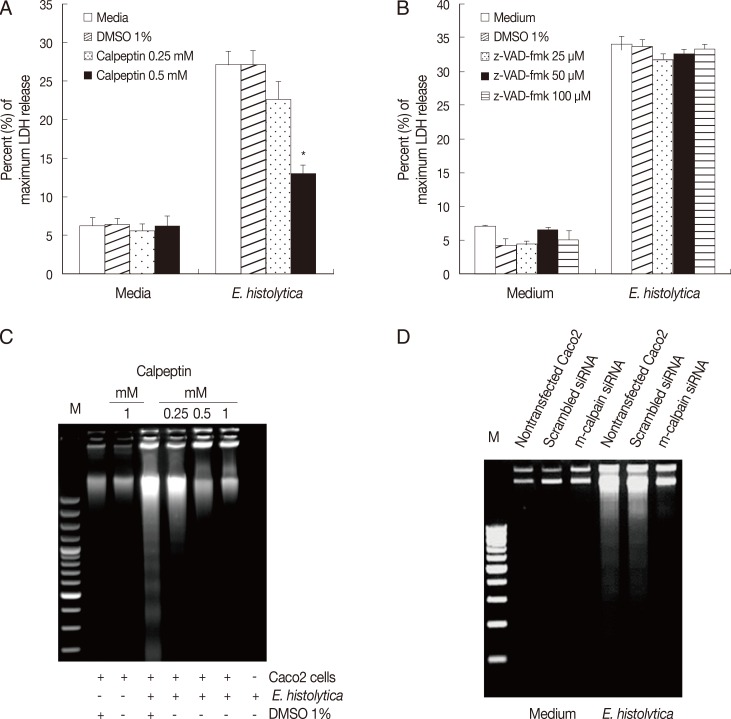
m-calpain is required for E. histolytica-induced cell death in Caco-2 cells. (A) Caco-2 cells (3×10⁵/sample) pretreated with either calpeptin (0.25-0.5 mM) or DMSO (1%) were incubated for 60 min with E. histolytica at a ratio of 5:1. Cell death was measured by the LDH release assay. Significant differences between groups are indicated as follows: *P<0.005. (B) Caco-2 cells (3×10⁵/sample) were incubatedfor 60 min with E. histolytica, at a ratio of 5:1, in either the absence or presence of z-VAD-fmk (25-100 mM). Cell death was measuredby the LDH release assay. Data are presented as means±SEM from three independent experiments. (C) Caco-2 cells (2.5×10⁶/sample) pretreated either with or without calpeptin (0.25-1 mM), or 1% DMSO (v/v) as a control, for 15 min at 37℃ were then incubated for 60 min at 37℃ in a CO2 incubator in either the absence or presence of E. histolytica (2.5×10⁵/sample). DNA fragmentation was subsequentlyanalysed by electrophoresis on 2% agarose gels. (D) At 72 hr post-transfection, Caco-2 cells were transfected with either scrambled siRNA (negative control) or m-calpain siRNA and subsequently co-incubated with E. histolytica prior to analysis of cell death by DNA fragmentation. Nontransfected Caco-2 cells served as an additional control.

