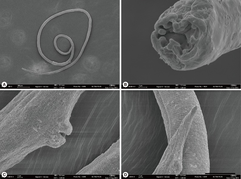
Fig. 4.
Scanning electron microscopic views of Strongyloides myopotami collected from the small intestine of a feral nutria. (A) Whole body view of a parasitic female. (B) En face view. 8-chambered type stoma with 8 circumoral elevations. (C) Prominent lips of vulva. (D) Posterior end showing a simple round tail.

