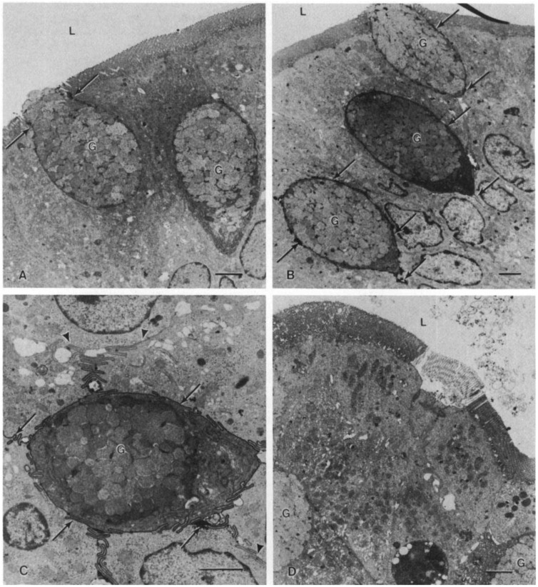
Figure 1.
Electron micrograph of isolated duodenal loops. A. Following exposure to normal saline, rare ruthenium red deposits could be seen around goblet cells (arrows). No deposits were visible between adjacent columnar epithelial cells (magnification of ×4600), B. Following aluminum citrate exposure, dense infiltration of ruthenium red deposits could be visualized around goblet cells (arrows). The mucosal epithelium following both normal saline and aluminum citrate was intact (magnification ×3700). C. A goblet cell, at higher magnification, following aluminum citrate treatment. Note the intense, ribbon-like outlining of the entire goblet cell’s intercellular space by ruthenium red which fades away at the junction of the columnar epithelial intercellular space (arrows) (magnification of ×7600). D. Aluminum chloride pre-incubation resulted in minimal or no ruthenium red in intercellular spaces but caused some patchy sloughing of mucosa (magnification of ×4400; G, goblet cell; L, lumen; bar represents 2 microns). Reprinted with permission from the International Society of Nephrology [16].

