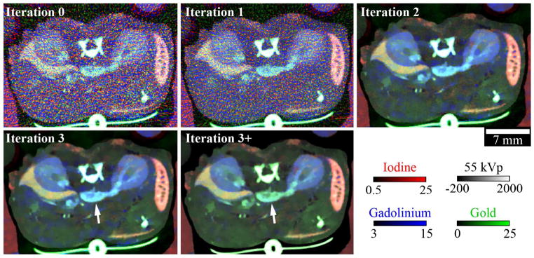
Figure 9. Spectral diffusion results by iteration.

Material maps overlaid on a single, 2D slice of the 55 kVp CT data after 0, 1, 2, and 3 iterations of spectral diffusion. “Iteration 3+” indicates the iteration 3 results after the application of subspace projection (Equation 2.27). White arrows indicate a blood vessel affected by subspace projection. The CT data and material maps are scaled as shown in HU (55 kVp data) and mg/mL (material maps).
