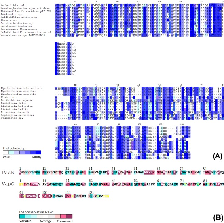
Figure 1.
(A) Multiple sequence alignment of VapC and PasB with most similar homologues and described family members. PasB (upper MSA) shows standard homology within RelE family (46% identity to the well-studied toxin from Escherichia coli). However, there are visible regions within active site that are unique and conserved among PasB like sequences. Strong homology can be observed for other non-related families, suggesting horizontal gene transfer. Color gradient corresponds to different amino acid hydrophobicity; (B) Conservation pattern calculated for protein families (for over 300 representatives).

