Figure 6.
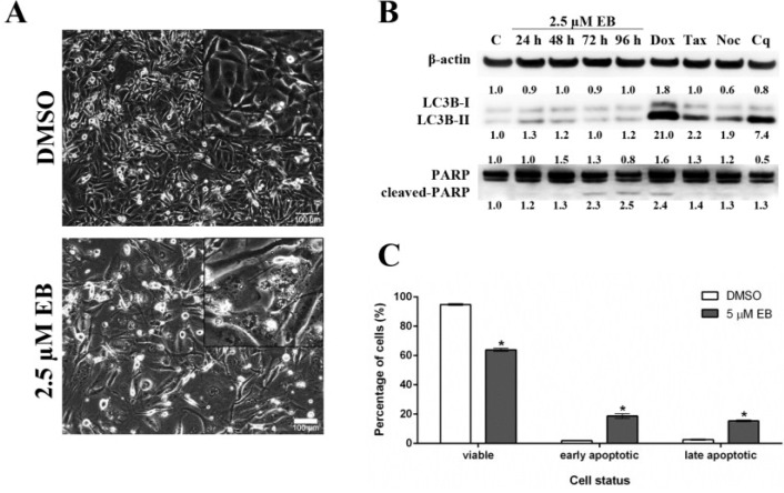
Eusynstyelamide B (EB, 1) induces cell death in MDA-MB-231 cells through apoptosis. (A) MDA-MB-231 cells were treated with 2.5 µM EB or 0.1% DMSO for 96 h and imaged using an Olympus IX70 microscope (10× objective, scale bar = 100 µm); (B) Cells were treated with 2.5 µM EB for the indicated times or 0.1% DMSO for 96 h (C). As positive controls for apoptosis (PARP cleavage), cell were treated with 1 µM doxorubicin (Dox) for 48 h, 2 nM taxol (Tax) for 24 h or 83 nM nocodazole (Noc) for 24 h. As a positive control for autophagy (LC3B-II), cell were treated with 25 µM chloroquine (Cq) for 48 h. Expression of the indicated proteins was assessed by Western blotting, normalized against the level of β-actin, and is expressed in fold-change relative to control (C); (C) MDA-MB-231 cells were treated with 5 µM EB or 0.1% DMSO for 96 h, stained with PI and Annexin V-FITC, and the number of viable, early apoptotic and late apoptotic/necrotic cells was quantified by flow cytometry (mean ± SD, n = 3, * p < 0.05).

