
Fig. 2.
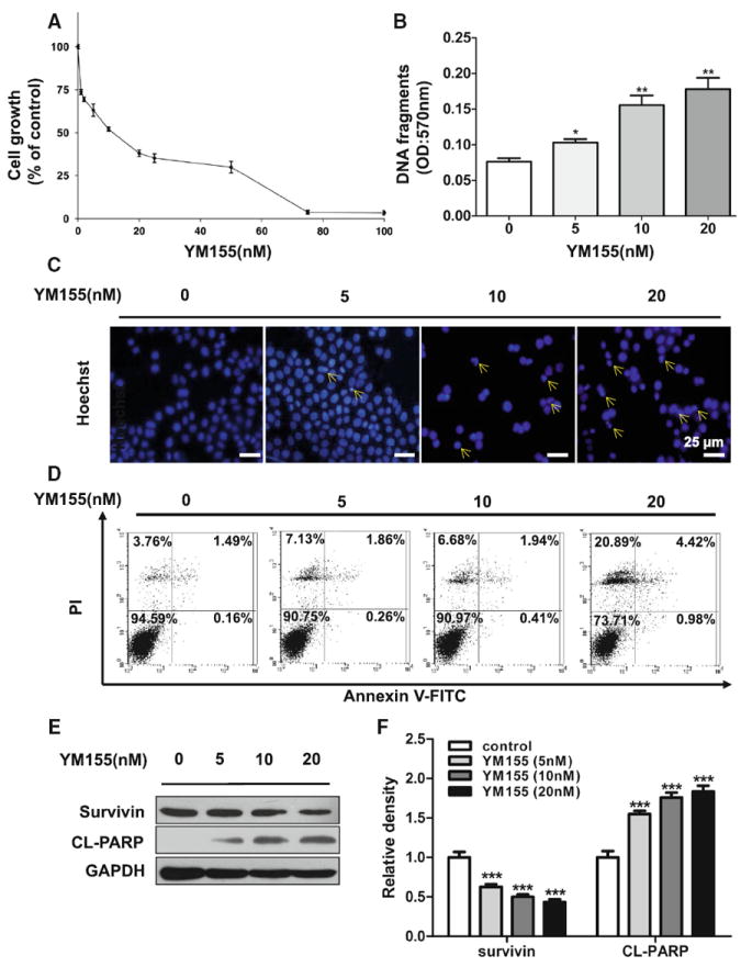
YM155 induced cell death in ACC-2 cell line. a Cell growth of ACC-2 was measured using an MTT assay after treated with YM155 for 24 h. The viability ratio is normalized by the ratio of the optical density value obtained from the YM155-treated sample divided by that of control group. b Cell death detection ELISA analysis of cell apoptosis induced by YM155 in dose-dependent manner in ACC-2 was detected. *P < 0.05; **P < 0.01 as compared with the control group. One-way ANOVA with post-Dunnett analysis was used. c The morphologic changes of ACC-2 treated with YM155 were captured using fluorescence microscopy with Hoechst 33258 staining. Scale bar 25 μm. d ACC cells were treated with YM155 for 24 h and apoptosis rate was determined by Annexin V-FITC/PI dual labeling using flow cytometry. e Cell lysates of the four groups subjected to western blotting with antibodies against survivin and cleaved PARP (CL-PARP), and GAPDH served as a loading control. f Relative quantitative data were calculated by ImageJ. ***P < 0.001 one-way ANOVA with post-Dunett analysis was used by GraphPad Prism5. The arrows indicate chromatin condensation in ACC-2 cells