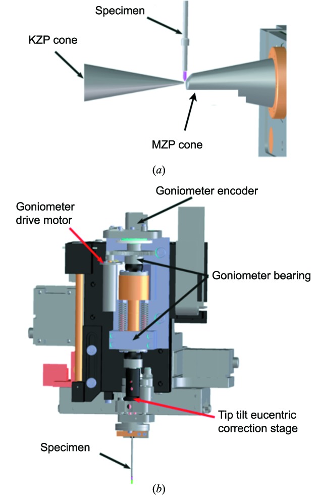
Figure 2.
(a) Close-up view of the specimen environment relative to the two zone plate cones. The specimen mounted in a thin-walled glass capillary is shown in pink [KZP = condenser zone plate; MZP = micro (objective) zone plate]. (b) Details of the specimen rotation stage, showing the relative position of the major components.

