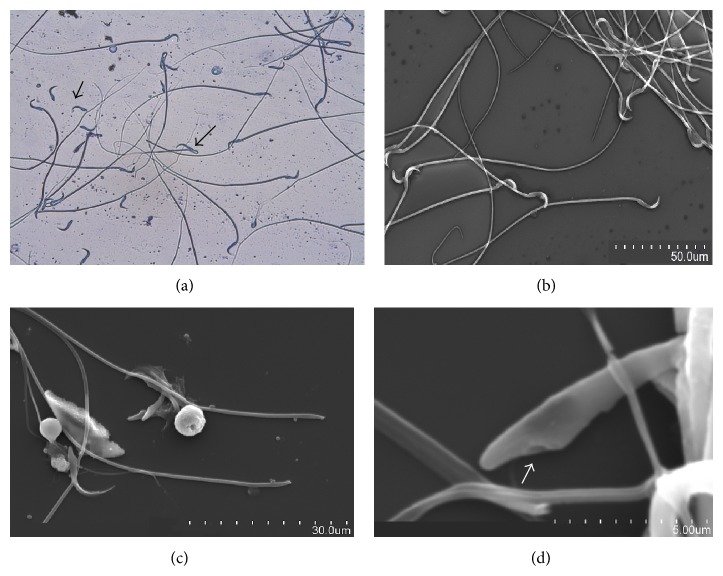
Figure 3.
(a–d) Cauda epididymal fluid smear of untreated rats and rats treated with triptolide showing sperm head tail separation. (a) Sperms with head tail separation (short arrow) and midpiece coiling (long arrow) observed in cauda epididymal fluid of treated rat under light microscope at 400x, (b) sperms with no head tail separation observed in cauda epididymal fluid of untreated rat under SEM at 15.0 kV 9.6 mm × 600 SE, (c) sperms with head tail separation observed in cauda epididymal fluid of treated rat under SEM at 15.0 kV 9.9 mm × 1.70 k SE, and (d) sperms with head tail separation observed in cauda epididymal fluid of treated rat under SEM at 15.0 kV 9.9 mm × 10 k SE. Arrow indicates region of separation of middle piece.

