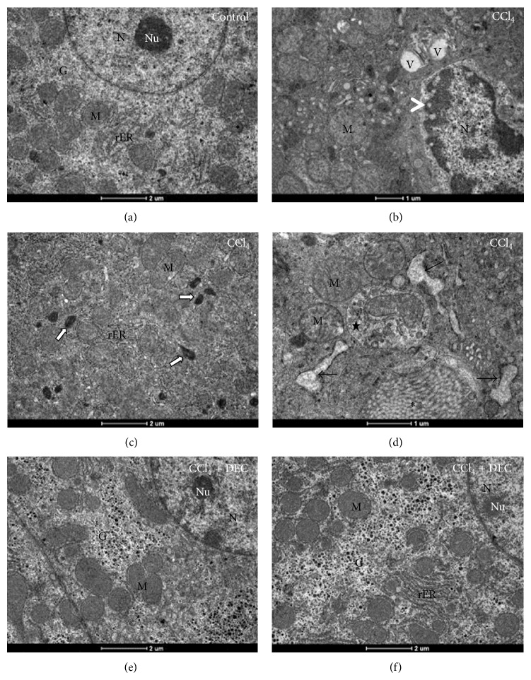
Figure 3.
Ultrathin sections of hepatocytes. (a) Control group; (b), (c) and (d) CCl4 group; (e) and (f) DEC + CCl4 group. Note that the chronic cell injury exhibits swollen mitochondria, several vacuoles (V) and lyses of chromatin, characteristic of a necrosis process (head arrows). Note also the collagen fibers (asterisk), rER dilatation (black arrows), and an autophagosome containing mitochondria (star). Mitochondria (M), glycogen (G), nucleus (N), nucleolus (Nu), rough endoplasmic reticulum (rER), and peroxisomes (white arrows). Bar: 1 and 2 μm.

