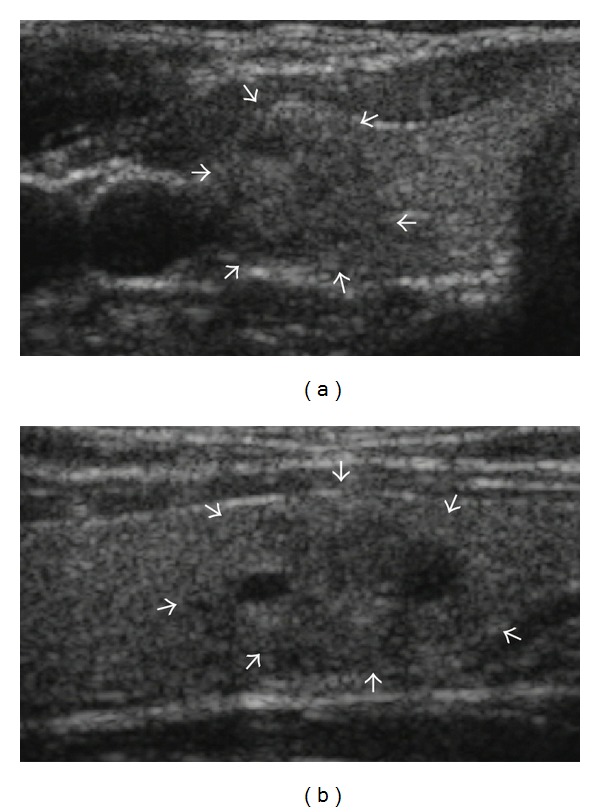
Figure 2.

Initially suspicious but concordant nodule after imaging-cytology correlation. US scans ((a) transverse; (b) longitudinal) in a 41-year-old female without remarkable medical history show a 16 mm sized predominantly solid mass (arrows) with microlobulated margin in the lower pole of the right lobe of the thyroid gland. The nodule was taller than wider on transverse scan. The initial cytologic result was adenomatous hyperplasia which was concordant with US findings considering relatively low PPV of these US finding in imaging-cytology correlation after biopsy. A follow-up US was recommended and nodule size gradually decreased from 16 mm to 13 mm with decrease of the cystic portion in follow-up US evaluations until July 2013 without any other significant changes in US features.
