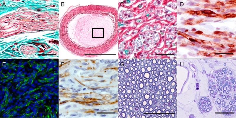
Figure 1.

Histological method as quality control in PNTE.
Longitudinal section of a peripheral nerve stained with Masson's trichrome method (A). Transversal section of a NeuraGen® collagen conduit stained with the MCOLL histochemical method with low (B) and high magnification (C), where it is possible to observe the collagen fibers in red, the myelin in blue and the nucleus darkly stained. Identification of Schwann cells with S-100, with the characteristic nuclear and cytoplasmatic positive reaction (D). Immunofluorescence and immunohistochemical identification of regenerated axons by using neurofilament (green) and GAP-43 (brown), respectively (E, F). Semithin transversal sections of native nerve (G) and regenerated nerve tissue (H) stained with toluidine blue. PNTE: Peripheral nerve tissue engineering; GAP-43: growth associated protein-43. Scale bar: 50 μm (A, D, F, G, and H), 100 μm (C, E) and 1 mm (B).
