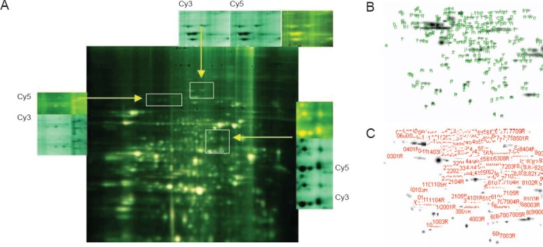
Figure 2.

Protein fingerprinting.
(A) The most obvious different spots are between I0minR0h and I25minR24h groups by paired experiments, we finally selected I25minR24h for electrophoresis (Cy3 and Cy5 fluorescence scanning). Cy3 and Cy5 are different fluorescent probes (B, C). Different protein spots were detected in the polyacrylamide gel by DeCyder software.
