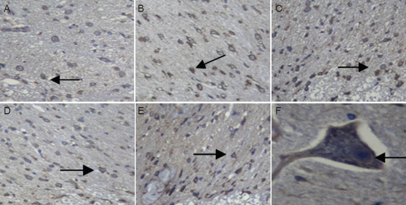
Figure 3.

Neurofilament protein M expression in rabbit spinal cord after ischemia/reperfusion (I/R) injury (immunohistochemistry).
(A) Sham group; (B) I25minR0 group; (C) I25minR12h group; (D) I25minR24h group; (E) I25minR48h group; (F) higher magnification image from the I25minR24h group showing neurofilament protein M expression in the neuronal membrane and axon. Arrows showed neurofilament protein M immunopositive cells. Original magnification: A–E, × 200; F, × 400.
