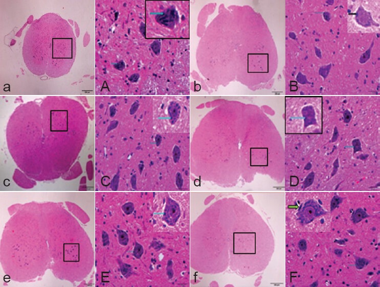
Figure 1.

Effect of intraperitoneal injection of ginsenoside Rd on the morphology of L2 spinal cord cross-sections in rats 5 days after spinal cord ischemia/reperfusion injury (hematoxylin-eosin staining).
(a, A) Sham group; (b, B) ischemia/reperfusion in-jury group (I/R group); (c, C) 6.25 mg/kg ginseno-side Rd group; (d, D) 12.5 mg/kg Rd group; (e, E) 25 mg/kg ginsenoside Rd group; (f, F) 50 mg/kg ginsenoside Rd group. (A) Large neurons, slightly stained nuclei, clearly visible nucleolus, and patchy Nissl bodies (arrows) were found in the cytoplasm. (B) Large neurons and slightly stained nuclei were found, the nucleolus was visible. Nissl body content (arrows) was significantly lower than that in the sham group. (C) The neurons were large; Nissl body content (arrows) was higher than that in the I/R group. (D) The neurons were large, with a large amount of cytoplasmic Nissl bodies (arrows). (E) The neurons were large and round, with slightly stained nuclei and thick nucleolus, there were a large number of Nissl bodies (arrows) in the cytoplasm. (F) The neurons were large and round, with slightly stained nuclei and thick nucleolus, and there were a large number of Nissl bodies (arrows) in the cytoplasm. Original magnification, a–f: × 40, A–F: × 400; A–F are the magnified images of the boxes in a–f, respectively.
