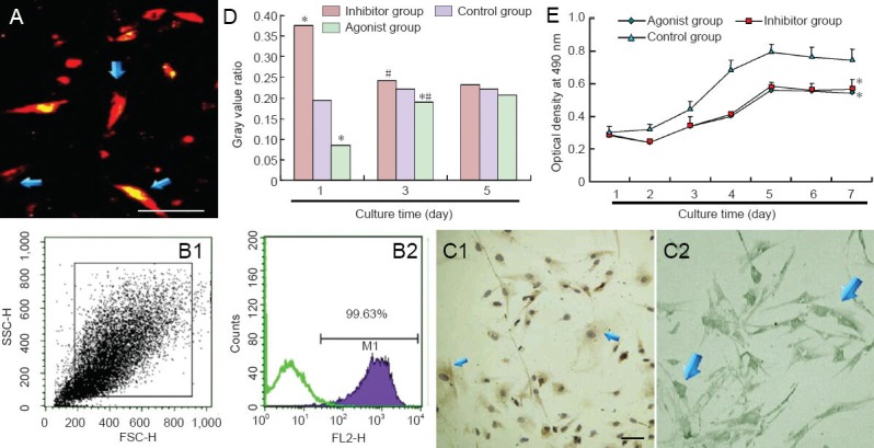
Figure 2.

Fluorescent labeling and pretreatment in bone marrow mesenchymal stem cells (BMSCs).
(A) CM-DiI-labeled BMSCs are red and spindle shaped. Scale bar: 50 μm. (B) Flow cytometry: Labeling rate of CM-DiI reached 99.63%. (C) Im-munohistochemical staining of protein phosphatase 2A (PP2A) (C1) and microtubule-associated protein 1B (MAP1B) (C2) expression in BMSC cytoplasm. Scale bar: 100 μm. (D) Effects of okadaic acid and N-acetyl-D-erythro-sphingosine on P1-MAP1B content in BMSCs (western blot assay). (E) Effects of okadaic acid and N-acetyl-D-erythro-sphingosine on BMSC viability. Data were expressed as the mean ± SD. Experiment was conducted in triplicate. *P < 0.05, vs. control group; #P < 0.05, vs. previous time point (one-way analysis of variance and independent samples t-test).
