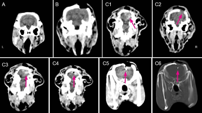
Figure 1.

CT images of rabbit skull at 5 hours after brain explosive injury.
(A) Blank control group: normal performance. (B) Sham surgery group: normal performance. (C) Explosive injury group: C1, subarachnoid hemorrhage (arrow); C2, brain tissue protrusion (arrow); C3, intracerebral hematoma (arrow); C4, intracranial pneumatocele (arrow); C5, residual iron (soft tissue window) (arrow), and C6 residual iron (bone window) (arrow). L: Left; R: right.
