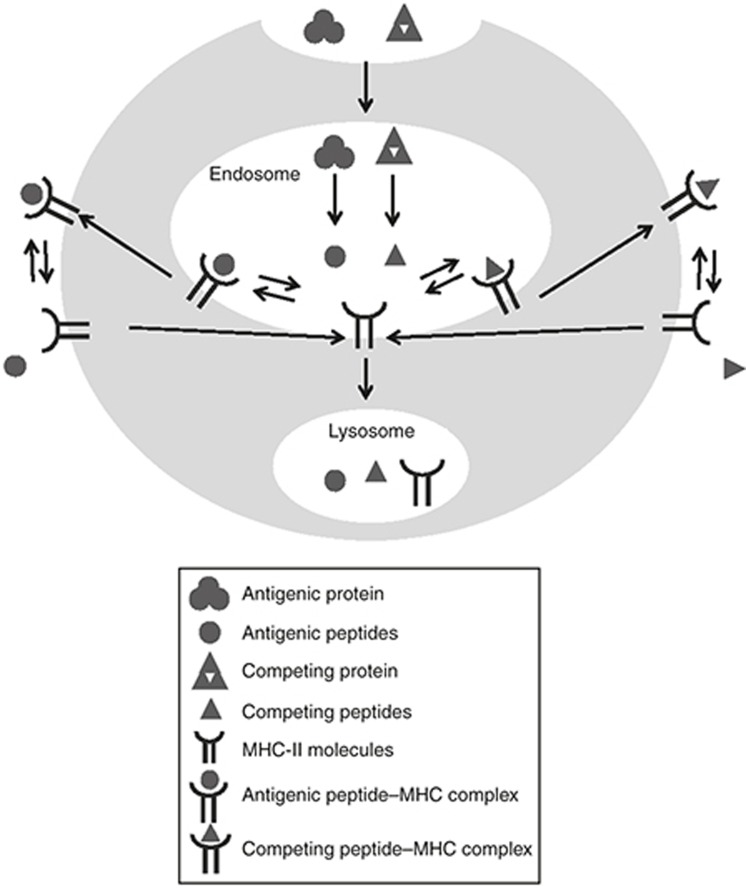
Figure 1.
Model structure for the subcellular level, including processes for antigen presentation in mature dendritic cells. The symbols in the figure legends are described below, with corresponding equation number in Supplementary Materials shown between parentheses.  : Antigenic protein, including antigenic protein in plasma (Ag, Eq. 27 in Supplementary Material) and antigenic protein in the endosome: (AgE, Eq. 4 in Supplementary Material);
  : antigenic peptide in endosome (pjE, Eq. 5 in Supplementary Material);
  : competing protein in the endosome (cpE, Eq. 9 in Supplementary Material);
  : competing peptide in the endosome (cptE, Eq. 10 in Supplementary Material);
  : MHC-II molecules, including those in the endosome (MEk, Eq. 6 in Supplementary Material) and those on dendritic cell membrane (Mk, Eq. 13 in Supplementary Material);
  : antigenic peptide-MHC complex, including those in the endosome (pjMEk, Eq. 7 in Supplementary Material) and those on cell membrane (pjMk, Eq. 8 in Supplementary Material);
  : competing peptide-MHC complex, including those in the endosome (cptMEk, Eq. 11 in Supplementary Material) and those on cell membrane (cptMk, Eq. 12 in Supplementary Material).

