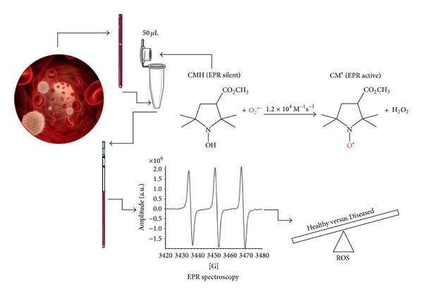
Figure 1.

EPR sample preparation and acquisition protocol. CMH Spin Probe (50 μL) is added in equal amount (1 : 1) to the collected capillary blood. The solution is immediately put in a glass EPR tube. From the generated radical compound, in the time course of the reaction, ten EPR spectra are collected in about 6 minutes, one of which is shown. The signal amplitude (a.u.) is proportional to the number of paramagnetic spin formed at the acquisition time. The calculated rate production values, converted in absolute levels (μmol·min−1) by using CP ∙ radical as external standard, allowed us to significantly discriminate the analyzed subjects' groups.
