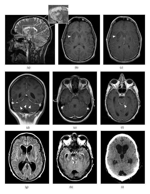
Figure 1.

Examples of abnormal neuroradiological findings in cryptococcal meningoencephalitis. (a) presents the typical pattern of cryptococcal gelatinous pseudocysts located in lentiform and caudate nuclei (T2WI hyperintense signal) while T1WI sequences ((b), arrow head) show a hypointense signal without enhancement in the T1 C+ image ((c), arrow head). Signs of meningitis are shown by leptomeningeal enhancement in the cerebellar Gyri in axial ((d), arrow heads) and coronal (e) T1 C+ scans. (f) presents signs of basal meningitis demonstrated by leptomeningeal enhancement (arrow head). Hydrocephalus is shown in axial FLAIR sequences in (g) and (h). The enlargement of the frontal and dorsal horns of the lateral ventricles with a hyperintense rim of the periventricular white matter indicates a CSF extravasation. A CCT scan performed 5 days later shows increased ballooning of the third ventricle and a progressive enlargement of the lateral ventricles (i).
