Figure 3.

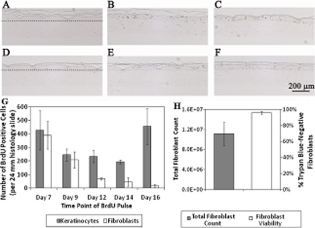
BrdU staining identifies proliferating cells in the BLCC throughout the culture process. BLCC medium remained unmodified (non-BrdU pulsed negative control) (A), or was pulsed with BrdU at Day 7 (B), Day 9 (C), Day 12 (D), Day 14 (E), or Day 16 (F), which was 12 hours prior to the scheduled media change. The scale bar in (F) represents 200 μm for (A), (B), (C), (D), (E), and (F). Dotted lines in (A) and (D) demarcate the epithelium–matrix interface. BrdU positive cells are stained brown. Images are representative of n = 3 constructs at each time point. Keratinocyte proliferation remained high up to Day 16, while fibroblast proliferation decreased over time (G). Proliferation was quantified (G) by counting the total number of BrdU-positive keratinocytes and fibroblasts per 24 mm biopsy sample (n = 3 samples per time point). Total fibroblast yield and viability were assessed at Day 20 (H) by collagenase digestion of the fibroblast matrix of the BLCC and trypan blue exclusion (n = 4). Data in (G) and (H) are presented as mean ± SD, n = 4. BLCC, bilayered living cellular construct; BrdU, bromodeoxyuridine.
