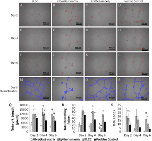
Figure 8.

BLCC and Epithelium only conditioned media promoted hUVEC network maintenance over the course of 6 days. Representative images at 5× magnification of hUVEC networks at Day 2 (A–D), Day 4 (E–H), and Day 6 (I–L) after application of conditioned media to preformed hUVEC networks on Matrigel show degradation (Fibroblast matrix) or maintenance (BLCC and Epithelium only) of the networks over time. Each image in A–L is a representative of n = 3 biological replicates and is a merge of phase contrast and fluorescence. Images were quantified using Wimasis software, and the output images from I–L are shown in M–P. Total network length (Q), total braching points (R), and total loops (S) are shown for each condition at all time points. For normally distributed tube length data, ANOVA with post hoc Tukey was performed. For nonnormally distributed branch point and total loop data, Kruskal–Wallis with pairwise mean rank comparisons was performed. *Indicates different from Fibroblast matrix and Epithelium only within same time point (p < 0.05). †Indicates different from Fibroblast matrix within same time point (p < 0.05). §Indicates different from Positive Control. Data are presented as mean ± SD, n = 3. BLCC, bilayered living cellular construct; hUVEC, human umbilical vein endothelial cell.
