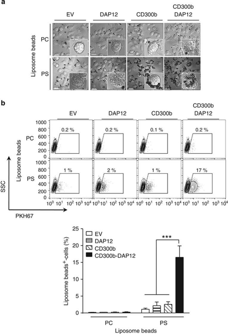
Figure 4.
CD300b-mediated engulfment of phosphatidylserine liposome-coated beads requires the co-expression of DAP12. (a) L929 cells transduced with the indicated constructs were incubated with PC- or PS-coated liposome beads for 20 min at 37 °C. The cells were fixed, and imaged by microscopy. The DIC images show cells and liposome-coated beads; the scale bar is 20 μm. The inserts show a close-up of a single cell. (b) PKH67-labeled L929 cells transduced with the indicated constructs were incubated with PC- or PS-coated liposome beads for 30 min. After homogenization of the cells, the beads were harvested, and their phagocytosis was determined by analysis of the percentage of PKH67+-beads using flow cytometry. The beads that became fluorescent were regarded as the engulfed beads, as they acquired the fluorescence due to their encapsulation within PKH67+-cell membranes (phagosomes). The contour plot indicates the gating strategy, and illustrates a representative result (upper panel). The bar graph (lower panel) shows the quantification of the percentage of engulfed liposome-coated beads. Data represent mean+S.E.M.; ***P<0.001 (ANOVA)

