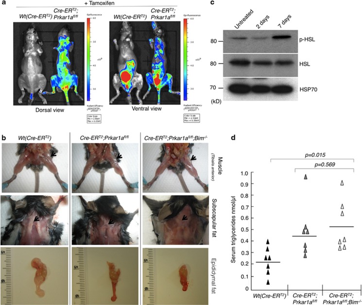
Figure 5.
Induced systemic deletion of Prkar1a caused cachexia in mice. (a) Imaging of mice undergoing cachexia with legumain (LE28) staining. In the ventral view, the control (ROSA-CreERT2) mouse also shows fluorescence in the bladder due to the clearance of the dye by the renal system. (b) Images of mice from the indicated genotypes showing loss of Tibialis anterior and subscapular and epididymal fat. (c) Western blot analysis of extracts of white adipose tissue from mice of the indicated genotypes 5 days after tamoxifen administration to determine the levels of activated hormone-induced lipase (p-HSL) and (d) serum triglyceride levels in mice measured (n=7 for each genotype)

