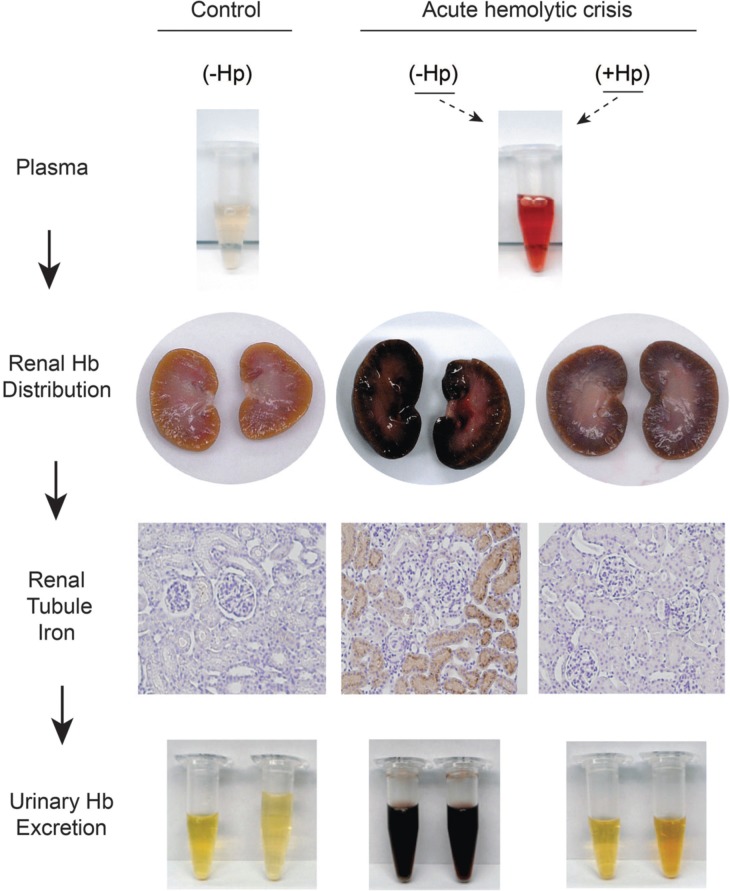
Figure 1.
Sequence of renal plasma clearance of hemoglobin: The sequence represents sustained intravascular hemolysis in a guinea pig model. The red color of Hb in plasma during hemolysis suggests ferrous Hb accumulation. In the presence of haptoglobin free Hb is sequestrated within the Hb-Hp complex. Processes of Hb-Hp clearance are saturated during massive hemolysis and the complex remains in circulation for an extended period of time, leading to the brownish color or ferric heme in bound Hb dimers, which is a result of auto-oxidation or reaction with NO or other oxidants in plasma. Free hemoglobin readily dissociates into dimers in plasma, leading to extensive glomerular filtration, which is blocked by Hp. The filtration of Hb leads to renal tubule iron deposition (brown) and hemoglobinuria. This is not observed in control animals or when Hp is administered and Hb remains sequestered in the Hb-Hp complex.

