Figure 2.

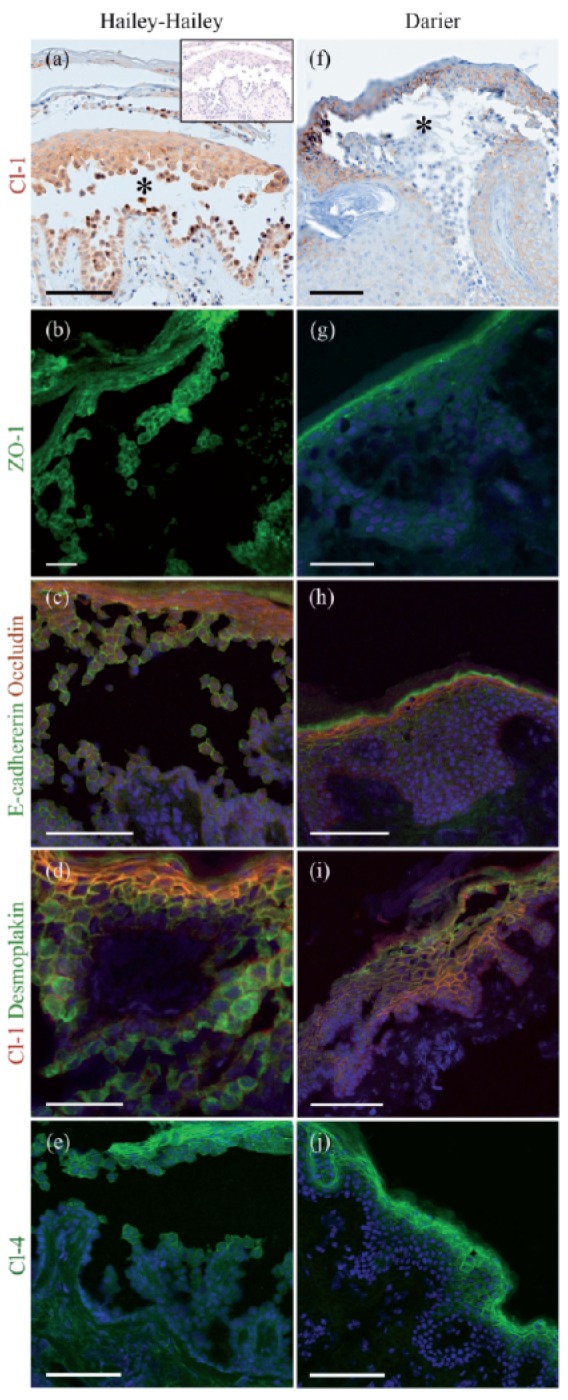
Hailey-Hailey disease (HHD) (a–e) and Darier's disease (DD) (f–j) lesions immunolabeled for tight junction proteins claudin-1 (a, f ), ZO-1 (b, g), and claudin-4 (e, j); double labeling for E-cadherin and occludin (c, h), and desmoplakin and claudin-1 (d, i). In (a) the inset is a control without any primary antibody. Avidin-biotin immunolabeling visualizes the suprabasal blisters (asterisks) in lesions of both diseases (a, f ). Claudin-1 is expressed in all epidermal cell layers (a, f). ZO-1 localizes to the upper epidermis and acantholytic cells in the HHD blister (b) while in DD, ZO-1 is expressed only in the granular cell layer (g). Occludin (red) is restricted to the granular cell layer in both diseases, while E-cadherin (green) is seen in intercellular junctions in all epidermal cell layers (c, h). Claudin-1 and desmoplakin are visible in all epidermal cell layers (d, i). Claudin-4 localizes to the upper epidermis and acantholytic cells in the Hailey-Hailey blister and in DD (e, j). Scale bar: 100 µm in (a), (c), (e), (f), (h), (i) and (j); 50 µm in (b), (d) and (g).
