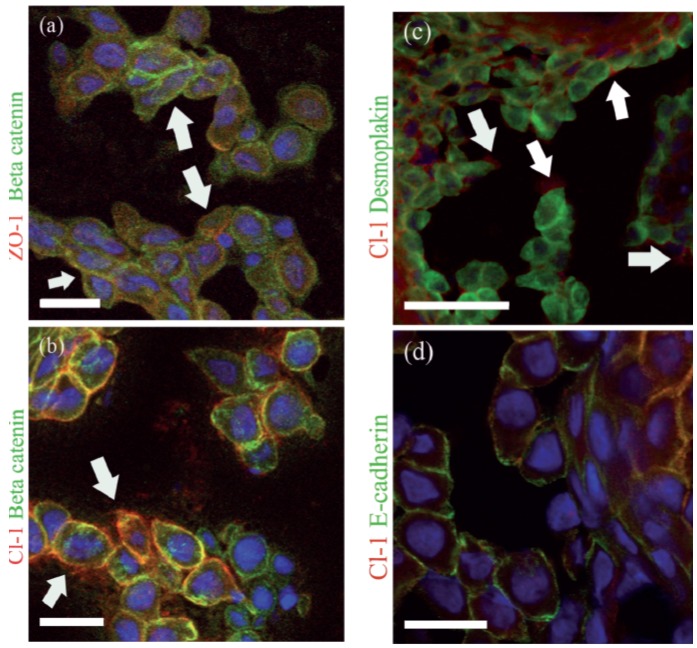
Figure 3.
Acantholytic cells in Hailey-Hailey disease (HHD) lesions, double immunolabeled for TJ proteins and adherens junction or desmosomal components. ZO-1 (red) and β-catenin (green) are present in some of the remaining intercellular junctions, while both proteins can be detected intracellularly and in plasma membranes not in contact with neighboring cells (arrows) (a). Claudin-1 and β-catenin in the cell-cell contacts (b). Claudin-1 is seen in the plasma membranes of cells not in contact with the neighboring cells (arrows) (b,c). Double labeling for desmoplakin (green) and claudin -1 (red) shows internalization of desmoplakin while claudin-1 stays in the plasma membrane (c). E-cadherin (green) and claudin-1 (red) are present at the plasma membrane (d). Scale-bar: 20 µm in (a), (b), (d); 50 µm in (c).

