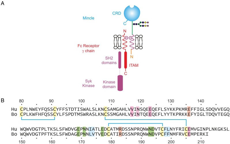
Fig. 1.
Organization of mincle. (A) Arrangement of mincle in the macrophage membrane, showing interactions with the common Fc receptor γ subunit within the lipid bilayer, which in turn interacts with Syk kinase on the cytoplasmic side of the membrane. ITAM, immunotyrosine activation motif. (B) Alignment of the amino acid sequences of the CRDs from human and bovine mincle. Disulfide bonds are indicated as cyan-colored lines between cysteine residues. Ligands for the principal Ca2+ are highlighted in green, ligands for the accessory Ca2+ are highlighted in violet, residues in the secondary sugar-binding site are highlighted in peach and residues that form the hydrophobic groove are highlighted in blue.

