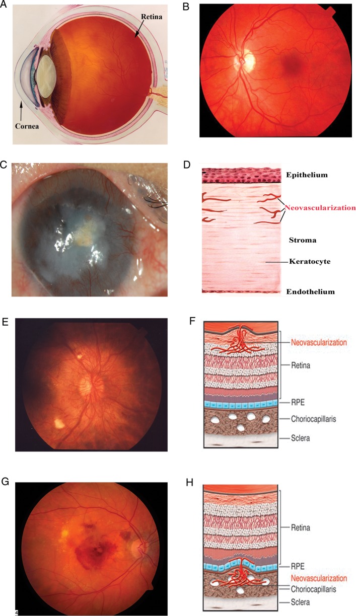
Fig. 1.
Schematic and photographic representation of the eye and corneal, retinal, and CNV. (A) Schematic depiction of the eye. (B) The fundus, i.e., the inner lining of a normal eye. (C and D) Normal cornea is transparent and avascular; in response to trauma, graft rejection or infection, blood vessels from the limbus (region where transparent cornea meets the opaque sclera) invade the cornea. (E and F) The retina is a highly ordered, multilayered structure that is richly vascularized. Diabetic retinopathy can lead to ischemia and neovascularization on the surface of the retina. (G and H) AMD can be associated with subretinal neovascularization originating from the choriocapillaris, and this can lead to subretinal hemorrhage. Credit: (A), (E) and (G) downloaded from National Eye Institute, NIH website with permission (ref.: NEA04, EDA01, EDA 24); (B) provided by J. S. Duker; (C) provided by Sugaya Satoshi and Tohru Sakimoto; (F) and (H) from Friedlander (2007) with permission.

