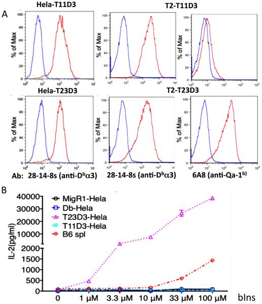
Fig. 3.
Expression of hybrid T11D3 and T23D3 molecules and test of the function of hybrid molecule expressing cells as Ag presenting cells. (A) FACS analysis of hybrid T11D3 and T23D3 on the surface of Hela and T2 cells. Transduced Hela cells were stained with 28-14-8s (α-Db α3) and T2 cells were stained with 28-14-8s and 6A8 (α-Qa-1b α2), respectively, shown as marked (red lines). The staining of isotype control Ab is blue. (B) Antigen presentation assay to test capability of the hybrid MHC-Ib molecule expressing cells to present insulin to 6C5 T hybridoma cells specific to bovine insulin (bINS). The assay was set up in triplicates (n=3) and the experiment was repeated twice with similar results. One of them was shown.

