
Fig. 6.
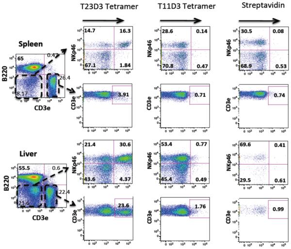
MHC Ib-Qdm tetramer staining of lymphocytes. B6 spleen and liver lymphocytes were stained with surface markers for B cells (B220), T cells (CD3ε), NK cells (NKp46) and the hybrid T23D3-Qdm or T11D3-Qdm tetramers. APC labeled streptavidin was used as a negative control. The lymphocytes were gated out for FACS analysis. The staining was repeated twice with similar results and one of each was shown.