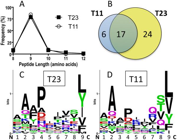
Fig. 7.

Peptide elution from Hela-T11D3 and Hela-T23D3 cells. (A) Length distribution of the peptides eluted from Hela-T11D3 and Hela-T23D3 cells. (B) The unique peptides from the T23 eluted and T11 eluted peptide pool were analyzed. Numbers of 8mer, 9mer and 10mer peptides were shown in the Venn diagram. (C) Sequence logo of the total eluted unique peptides from T23 and (D) T11. Each column represents one amino acid position in the peptide. Amino acids with different properties were labeled with different colors.
