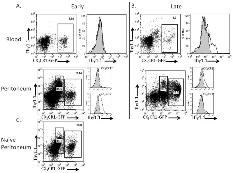
Figure 3. IL-10 production is initiated in CX3CR1-negative MDSCs and subsequently is detected in CX3CR1 -positive MDSCs in tumor-bearing mice.
Blood and peritoneal samples from 2 (A) and 4-week (B) ID8vβ tumor-bearing mice were assessed for IL-10 reporter staining. Plots of CX3CR1-GFP/IL-10BiT reporter staining and histograms indicate reporter staining in gated populations in CX3CR1-GFP/IL-10BiT mice (black) in comparison to CX3CR1-GFP control mice (gray, filled) representative of results from ≥5 mice. (C) Staining from the peritoneum of naïve CX3CR1-GFP/IL-10BiT reporter.

