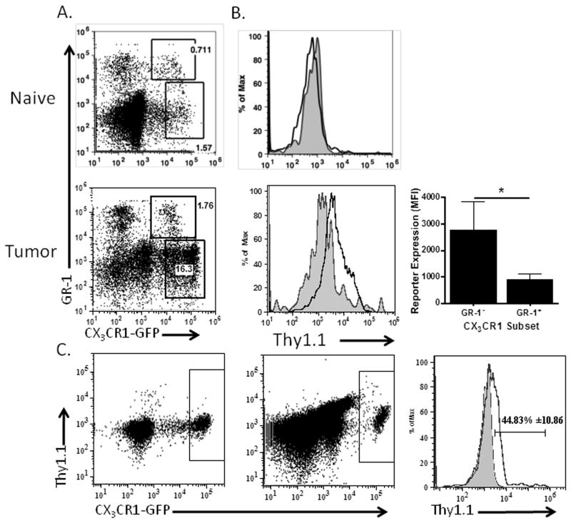
Figure 5. IL-10 reporter is induced in infiltrating CX3CR1-positive monocytic MDSC.
(A) GR-1 positive and negative CX3CR1-positive cells in the naïve peritoneum, or ascites of tumor-bearing CX3CR1-GFP/IL-10BiT mice were analyzed for IL-10 reporter staining. Plot shows GR-1 positive and negative gates. (B) Histogram indicates reporter staining on gated populations from CX3CR1-GFP/IL-10BiT mice showing GR-1 negative (black) compared to GR-1 positive (gray, filled) with quantification of IL-10 reporter staining from gated populations (panel A) by MFI. (C) Plots demonstrating reporter staining on CX3CR1-GFP positive cells before (left dot plot) and after transfer (right dot plot) of PBMC from CX3CR1-GFP/IL-10BiT mice i.p. into a wild-type tumor bearing recipient, with comparative histogram of IL-10 reporter staining pre (gray filled) and post (black) transfer showing percentage positive for reporter staining with standard deviation. n=3 independent experiments, 3 mice. Statistical significance (*p < 0.05) and standard deviations shown.

