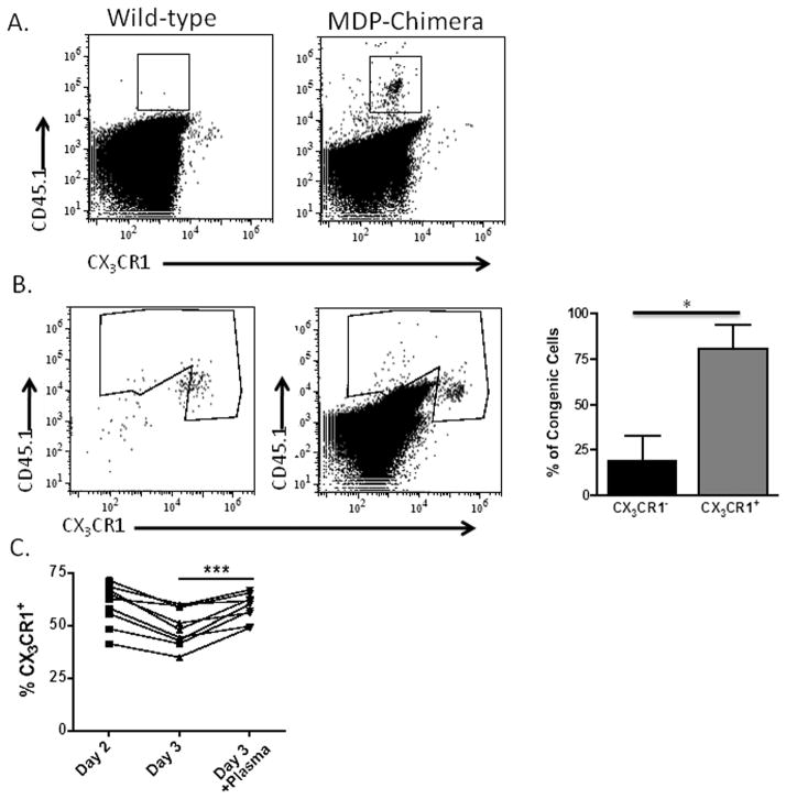
Figure 6. Tumor-infiltrating myeloid cells generated from the macrophage/dendritic cell precursor (MDP) lose CX3CR1 expression, but tumor-transferred monocytes maintain CX3CR1 expression.
(A) Ascites from ID8vβ tumor-bearing wild-type mouse (left) and MDP-reconstituted (right) mouse were assessed for CX3CR1-negative (gate shown), congenically-marked cells results representative of 2 independent experiments, 6 mice. (B) Sorted CX3CR1-GFP positive cells from naïve blood (left plot) were adoptively transferred into the peritoneum of tumor bearing wild-type mice and assessed for GFP expression 4 days post-transfer (right plot) with quantification of CX3CR1 positive and negative subsets in the congenic gate with standard deviation (n=4, 4 mice). (C) Percentages of CX3CR1-positive cells pre-gated for CD11b+Ly6G− in bone marrow cultured overnight, and with our without addition of tumor ascites plasma for an additional 24 hours (n=9, 3 independent experiments). Statistical significance (***p < 0.001 and *p<.05) and standard deviations shown.

