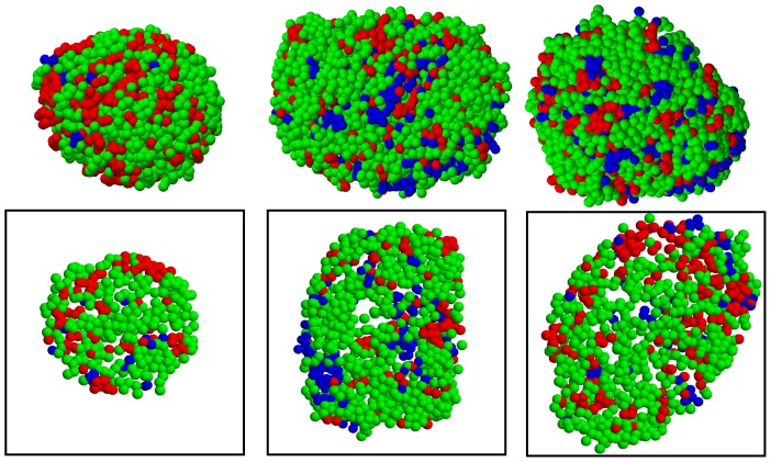
Figure 8. Cellular organization of human pancreatic islets.
Three-dimensional spatial distribution of  cells (red),
  cells (green), and
  cells (blue) in human islets is shown. To show internal islet structures clearly, their corresponding two-dimensional sections are also shown in boxes. Note that islets are isolated from the Human3 subject for this plot.

